Signs and symptoms | Diagnosis | Classification | Treatment | Children
A bone fracture (sometimes abbreviated FRX or Fx, Fx, or #) is a medical condition in which there is a damage in the continuity of the bone. A bone fracture can be the result of high force impact or stress, or a minimal trauma injury as a result of certain medical conditions that weaken the bones, such as osteoporosis, bone cancer, or osteogenesis imperfecta, where the fracture is then properly termed a pathologic fracture.
Although broken bone and bone break are common colloquialisms for a bone fracture, break is not a formal orthopedic term.
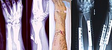
Internal and external views of an arm with a compound fracture, both before and after surgery.
|
Signs and symptoms
Although bone tissue itself contains no nociceptors, bone fracture is painful for several reasons:
- Breaking in the continuity of the periosteum, with or without similar discontinuity in endosteum, as both contain multiple pain receptors.
- Edema of nearby soft tissues caused by bleeding of torn periosteal blood vessels evokes pressure pain.
- Muscle spasms trying to hold bone fragments in place. Sometimes also followed by cramping.
Damage to adjacent structures such as nerves or vessels, spinal cord and nerve roots (for spine fractures), or cranial contents (for skull fractures) can cause other specific signs and symptoms.
Effects of smoking
Smokers generally have lower bone density than non-smokers, so have a much higher risk of fractures. There is also evidence that smoking delays bone healing. Some research indicates, for example, that it delays tibial shaft fracture healing from a median healing time of 136 to 269 days.[5] This means that the fracture healing time was approximately doubled in smokers. Although some other studies show less extreme effects, it is still shown that smoking delays fracture healing.
Diagnosis

Radiography to identify possible fractures after a knee injury.
A bone fracture may be diagnosed based on the history given and the physical examination performed. Radiographic imaging is often performed, to confirm the diagnosis. Under certain circumstances, radiographic examination of the nearby joints is indicated in order to exclude dislocations and fracture-dislocations. In situations where projectional radiography alone is insufficient, Computed Tomography (CT) or Magnetic Resonance Imaging (MRI) may be indicated.
Classification
"Compound Fracture" redirects here. For the 2013 horror film, see Compound Fracture (film).
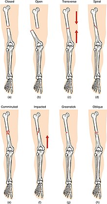
Compare healthy bone with different types of fractures:
(a) closed fracture
(b) open fracture
(c) transverse fracture
(d) spiral fracture
(e) comminuted fracture
(f) impacted fracture
(g) greenstick fracture
(h) oblique fracture

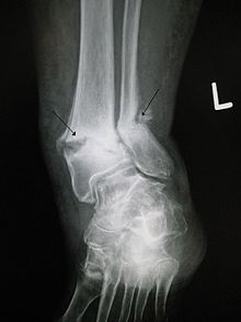
Open ankle fracture with luxation
In orthopedic medicine, fractures are classified in various ways. Historically they are named after the doctor who first described the fracture conditions. However, there are more systematic classifications in place currently.
Mechanism
- Traumatic fracture - This is a fracture due to sustained trauma. e.g.- Fractures caused by a fall, road traffic accident, fight etc.
- Pathologic fracture - A fracture through a bone which has been made weak by some underlying disease is called pathological fracture. e.g.- a fracture through a bone weakened by metastasis. Osteoporosis is the most common cause of pathological fracture.
- Periprosthetic fracture - A fracture at the point of mechanical weakness at the end of an implant
Soft-tissue involvement
- Closed fracture: are those in which the overlying skin is intact
- Open fracture/Compound fracture: involve wounds that communicate with the fracture, or where fracture hematoma is exposed, and may thus expose bone to contamination. Open injuries carry a higher risk of infection.
- Clean fracture
- Contaminated fracture
Displacement
- Non-displaced
- Displaced
- Translated
- Angulated
- Rotated
- Shortened
Fracture pattern
- Linear fracture: A fracture that is parallel to the bone's long axis.
- Transverse fracture: A fracture that is at a right angle to the bone's long axis .
- Oblique fracture: A fracture that is diagonal to a bone's long axis (more than 30°).
- Spiral fracture: A fracture where at least one part of the bone has been twisted.
- Compression fracture/Wedge fracture: usually occurs in the vertebrae, for example when the front portion of a vertebra in the spine collapses due to osteoporosis (a medical condition which causes bones to become brittle and susceptible to fracture, with or without trauma).
- Impacted fracture: A fracture caused when bone fragments are driven into each other.
- Avulsion fracture: A fracture where a fragment of bone is separated from the main mass.
Fragments
- Incomplete fracture: A fracture in which the bone fragments are still partially joined. In such cases, there is a crack in the osseous tissue that does not completely traverse the width of the bone
- Complete fracture: A fracture in which bone fragments separate completely.
- Comminuted fracture: A fracture in which the bone has broken into several pieces.
Anatomical location
An anatomical classification may begin with specifying the involved body part, such as the head or arm, followed with more specific localization. Fractures that have additional definition criteria than merely localization can often be classified as subtypes of fractures that merely are, such as a Holstein-Lewis fracture being a subtype of a humerus fracture. However, most typical examples in an orthopedic classification given in previous section cannot appropriately be classified into any specific part of an anatomical classification, as they may apply to multiple anatomical fracture sites.
- Skull fracture
- Basilar skull fracture
- Blowout fracture - a fracture of the walls or floor of the orbit
- Mandibular fracture
- Nasal fracture
- Le Fort fracture of skull - facial fractures involving the maxillary bone and surrounding structures in a usually bilateral and either horizontal, pyramidal or transverse way.
- Spinal fracture
- Cervical fracture
- Fracture of C1, including Jefferson fracture
- Fracture of C2, including Hangman's fracture
- Flexion teardrop fracture - a fracture of the anteroinferior aspect of a cervical vertebral
- Clay-shoveler fracture - fracture through the spinous process of a vertebra occurring at any of the lower cervical or upper thoracic vertebrae
- Burst fracture - in which a vertebra breaks from a high-energy axial load
- Compression fracture - a collapse of a vertebra, often in the form of wedge fractures due to larger compression anteriorly.
- Chance fracture - compression injury to the anterior portion of a vertebral body with concomitant distraction injury to posterior elements
- Holdsworth fracture - an unstable fracture dislocation of the thoracolumbar junction of the spine
- Rib fracture
- Sternal fracture
- Shoulder fracture
- Clavicle fracture
- Scapular fracture
- Arm fracture
- Humerus fracture (fracture of upper arm)
- Supracondylar fracture
- Holstein-Lewis fracture - a fracture of the distal third of the humerus resulting in entrapment of the radial nerve.
- Forearm fracture
- Ulnar fracture
- Monteggia fracture - a fracture of the proximal third of the ulna with the dislocation of the head of the radius
- Hume fracture - a fracture of the olecranon with an associated anterior dislocation of the radial head
- Radius fracture
- Essex-Lopresti fracture - a fracture of the radial head with concomitant dislocation of the distal radio-ulnar joint with disruption of the interosseous membrane.[6]
- Distal radius fracture
- Galeazzi fracture - a fracture of the radius with dislocation of the distal radioulnar joint
- Colles' fracture - a distal fracture of the radius with dorsal (posterior) displacement of the wrist and hand
- Smith's fracture - a distal fracture of the radius with volar (ventral) displacement of the wrist and hand
- Barton's fracture - an intra-articular fracture of the distal radius with dislocation of the radiocarpal joint.
- Hand fracture
- Scaphoid fracture
- Rolando fracture - a comminuted intra-articular fracture through the base of the first metacarpal bone
- Bennett's fracture - a fracture of the base of the first metacarpal bone which extends into the carpometacarpal (CMC) joint.
- Boxer's fracture - a fracture at the neck of a metacarpal
- Pelvic fracture
- Fracture of the hip bone
- Duverney fracture - an isolated pelvic fracture involving only the iliac wing.
- Femoral fracture
- Hip fracture (anatomically a fracture of the femur bone and not the hip bone)
- Patella fracture
- Crus fracture
- Tibia fracture
- Pilon fracture
- Tibial plateau fracture
- Bumper fracture - a fracture of the lateral tibial plateau caused by a forced valgus applied to the knee
- Segond fracture - an avulsion fracture of the lateral tibial condyle
- Gosselin fracture - a fractures of the tibial plafond into anterior and posterior fragments
- Toddler's fracture - an undisplaced and spiral fracture of the distal third to distal half of the tibia
- Fibular fracture
- Maisonneuve fracture - a spiral fracture of the proximal third of the fibula associated with a tear of the distal tibiofibular syndesmosis and the interosseous membrane.
- Le Fort fracture of ankle - a vertical fracture of the antero-medial part of the distal fibula with avulsion of the anterior tibiofibular ligament.
- Bosworth fracture - a fracture with an associated fixed posterior dislocation of the proximal fibular fragment which becomes trapped behind the posterior tibial tubercle. The injury is caused by severe external rotation of the ankle.
- Combined tibia and fibula fracture
- Trimalleolar fracture - involving the lateral malleolus, medial malleolus and the distal posterior aspect of the tibia
- Bimalleolar fracture - involving the lateral malleolus and the medial malleolus.
- Pott's fracture
- Foot fracture
- Lisfranc fracture - in which one or all of the metatarsals are displaced from the tarsus
- Jones fracture - a fracture of the proximal end of the fifth metatarsal
- March fracture - a fracture of the distal third of one of the metatarsals occurring because of recurrent stress
- Calcaneal fracture
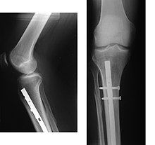
X-ray showing the proximal portion of a fractured tibia with anintramedullary nail.
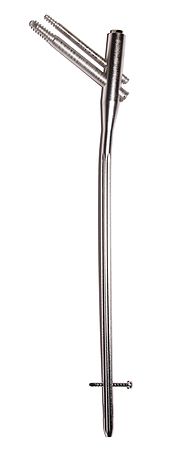
Proximal femur nail with locking and stabilisation screws for treatment of femur fractures of left thigh.

The surgical treatment ofmandibular angle fracture. Fixation of the bone fragments by the plates. The principles of osteosynthesis are stability (immobility of the fragments that creates the conditions for bones coalescence) and functionality.
Treatment of bone fractures are broadly classified as surgical or conservative, the latter basically referring to any non-surgical procedure, such as pain management, immobilization or other non-surgical stabilization. A similar classification is open versus closed treatment, in which open treatment refers to any treatment in which the fracture site is surgically opened, regardless of whether the fracture itself is anopen or closed fracture.
Pain management
In arm fractures in children, ibuprofen has been found to be as effective as a combination of acetaminophen and codeine.
Immobilization
Since bone healing is a natural process which will most often occur, fracture treatment aims to ensure the best possible function of the injured part after healing. Bone fractures are typically treated by restoring the fractured pieces of bone to their natural positions (if necessary), and maintaining those positions while the bone heals. Often, aligning the bone, called reduction, in good position and verifying the improved alignment with an X-ray is all that is needed. This process is extremely painful without anesthesia, about as painful as breaking the bone itself. To this end, a fractured limb is usually immobilized with a plaster or fiberglass cast or splint which holds the bones in position and immobilizes the joints above and below the fracture. When the initial post-fracture edema or swelling goes down, the fracture may be placed in a removable brace or orthosis. If being treated with surgery, surgical nails, screws, plates and wires are used to hold the fractured bone together more directly. Alternatively, fractured bones may be treated by the Ilizarov method which is a form of external fixator.
Occasionally smaller bones, such as phalanges of the toes and fingers, may be treated without the cast, by buddy wrapping them, which serves a similar function to making a cast. By allowing only limited movement, fixation helps preserve anatomical alignment while enabling callus formation, towards the target of achieving union.
Splinting results in the same outcome as casting in children who have a distal radius fracture with little shifting.
Surgery
Surgical methods of treating fractures have their own risks and benefits, but usually surgery is done only if conservative treatment has failed, is very likely to fail, or likely to result in a poor functional outcome. With some fractures such as hip fractures (usually caused by osteoporosis), surgery is offered routinely because non-operative treatment results in prolonged immobilisation, which commonly results in complications including chest infections, pressure sores, deconditioning, deep vein thrombosis (DVT) and pulmonary embolism, which are more dangerous than surgery. When a joint surface is damaged by a fracture, surgery is also commonly recommended to make an accurate anatomical reduction and restore the smoothness of the joint.
Infection is especially dangerous in bones, due to the recrudescent nature of bone infections. Bone tissue is predominantly extracellular matrix, rather than living cells, and the few blood vessels needed to support this low metabolism are only able to bring a limited number of immune cellsto an injury to fight infection. For this reason, open fractures and osteotomies call for very careful antiseptic procedures and prophylacticantibiotics.
Occasionally bone grafting is used to treat a fracture.
Sometimes bones are reinforced with metal. These implants must be designed and installed with care. Stress shielding occurs when plates or screws carry too large of a portion of the bone's load, causing atrophy. This problem is reduced, but not eliminated, by the use of low-modulusmaterials, including titanium and its alloys. The heat generated by the friction of installing hardware can easily accumulate and damage bone tissue, reducing the strength of the connections. If dissimilar metals are installed in contact with one another (i.e., a titanium plate withcobalt-chromium alloy or stainless steel screws), galvanic corrosion will result. The metal ions produced can damage the bone locally and may cause systemic effects as well.
Electrical bone growth stimulation or osteostimulation has been attempted to speed or improve bone healing. Results however do not support its effectiveness.
Complications
An old fracture with nonunion of the fracture fragments.
Some fractures can lead to serious complications including a condition known as compartment syndrome. If not treated, compartment syndrome can eventually require amputation of the affected limb. Other complications may include non-union, where the fractured bone fails to heal or mal-union, where the fractured bone heals in a deformed manner.
Complications of fractures can be classified into three broad groups depending upon their time of occurrence. These are as follows -
- Immediate complications - occurs at the time of the fracture.
- Early complications - occurring in the initial few days after the fracture.
- Late complications - occurring a long time after the fracture.
Children
In children, whose bones are still developing, there are risks of either a growth plate injury or a greenstick fracture.
- A greenstick fracture occurs due to mechanical failure on the tension side. That is, since the bone is not as brittle as it would be in an adult, it does not completely fracture, but rather exhibits bowing without complete disruption of the bone's cortex in the surface opposite the applied force.
- Growth plate injuries, as in Salter-Harris fractures, require careful treatment and accurate reduction to make sure that the bone continues to grow normally.
- Plastic deformation of the bone, in which the bone permanently bends but does not break, is also possible in children. These injuries may require an osteotomy (bone cut) to realign the bone if it is fixed and cannot be realigned by closed methods.
- Certain fractures are known to occur mainly in the pediatric age group, such as fracture of the clavicle & supracondylar fracture of the humerus.
Back to Top


